


ABOUT
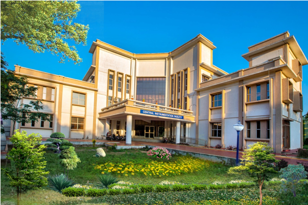
Research excellence in Science and Technology of an institution is increasingly dependent on the availability of advanced instrumental facilities and easy accessibility. The idea of establishing a Central Instrumental Facility (CIF) with high-end equipment under one roof saw the light when Honourable Chief Minister of Odisha Shri Naveen Patnaik declared to provide funds to establish a Central Instrumental Facility at Utkal University. A dedicated three-storeyed CIF building was built at a central place in the University campus with the aid of the Government of Odisha.
The vision of the CIF is to provide a central facility with the latest and advanced analytical techniques for cutting-edge research in various areas of science and technology and has set the following objectives:
- To provide facilities for sophisticated analytical instruments to facilitate multidisciplinary research and to cater to the needs of the academic departments of the University.
- To extend the facility to the researchers from other institutes as and when required.
- To provide guidance for the acquisition of data using sophisticated Instruments.
- To organise short-term courses/workshops/training programmes on the use and applications of various analytical techniques for students, research scholars, laboratory assistants and faculty of Utkal University and also from other Laboratories, Universities and Industries.
- To augment the instrumental facility from time to time to establish the centre as a major facility for spectral measurements, molecular and crystal structure determination, and imaging analyses in the eastern region.
Advisory Committee
- Chairperson, Post Graduate Council, Utkal University
- Director, Research & Development, Utkal University
- Coordinator, IDP, WB-OHEPEE, Utkal University
- Director, Institute of Physics, Bhubaneswar
- Director, NISER, Bhubaneswar
- B. R. Nayak, Principal Scientist, CSIR-IMMT, Bhubaneswar
- Director, Institute of Life Sciences, Bhubaneswar
- Sashikanta Dash, Dy. Director, Department of Biotechnology, DST, Govt. of Odisha, Bhubaneswar
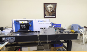
MICRO RAMAN SPECTROMETER
Labram HR Evo, Raman Spectroscopy, LASER Source: 532 nm, 633 nm and 325 nm
Raman spectroscopy is a molecular spectroscopic technique that makes use of the interaction between light and matter to shed light on the composition or properties of a material.
In contrast to IR spectroscopy, which relies on light absorption, Raman spectroscopy obtains its information through the process of light dispersion. Raman spectroscopy can add to our understanding of a reaction by revealing information about intra- and intermolecular vibrations. Raman spectroscopy can be used to determine a substance’s identity since it produces a spectrum that is representative of the precise vibrations of a certain molecule (a “molecular fingerprint”). Additional details regarding lower frequency modes and vibrations that reveal details of the crystal lattice and molecule backbone structure can be obtained using Raman spectroscopy.
HPC PARAM SHAVAK: 2
(Intel Xeon Process with 14 cores each having 2.4 GHz clock speed) Installed at the Computer Centre of Utkal University as a Central Facility.
HPC @ COE, HECMP
CoE in High Energy and Condensed Matter Physics has a High Performance Computing Facility (HPC) with 20 nodes, Intel processors, Intel® C621 Series Chipset Motherboard with storage capacity 144 TB (12 X 12TB) 7.2K RPM.

It has the capacity to perform most demanding computational work especially for research problems that involve large data sets, molecular dynamics and numerical calculations. It provides advanced computational facilities to the students and faculties of Utkal University. It supports many freeware as well as paid software including numerical packages, libraries and C and Fortran compilers to enhance academic scientific research. MedeA-VASP simulation software for first-principles calculations and Gaussian with TCP Linda Support & GaussView for computational chemistry. The facility is available for 24/7, 365 days to university users to maintain high academic standards in teaching and research at the international level.
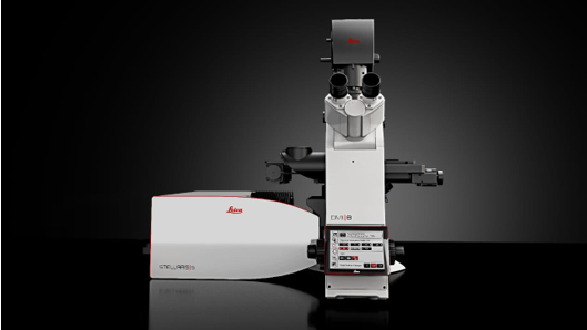
CONFOCAL MICROSCOPE WITH LIVE CELL IMAGING FACILITY (Under Installation)
Make: Leica, Model: Srellaris 5 LIAchroic with accessories
Confocal microscopy is a powerful technique used to create 3D images of the structures within living cells and to examine the dynamics of cellular processes. For high-speed imaging of fluorescent molecules and structures within living cells, microscopes must provide rapid field-of-view scanning that eliminates any out-of-focus light planes that can obscure fluorescence.
- Imaging dynamic microphometric changes in cells and organ cultures using fluorescence markers to understand properties such as cell growth, differentiation and migration.
- Photo activation or photo-conversion experiments to mark a single cell, organelles in a cell or molecules in a cell to track dynamic changes over time in response to changes in environment.
- Intracellular trafficking such as cargo delivery and their fate using fluorescence probes.
- Quantifying the proximity and interaction molecules in a living cell.
- Imaging of dynamic changes in the cellular physiology such as changes in the levels of calcium, pH, etc.
- Checking efficiency of contrast reagents and organ cultures.
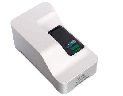
PARTICLE SIZE ZETA POTENTIAL ANALYSER (Under Installation)
Make: Melverl Panalytical, Model: ZetaSizer Advanced Pro Blue
This equipment measures a wide range of sample types and concentration. It offers wide dynamic concentration capability and high sensitivity due to non-invasive back seatter (NIBS) optical design. Measurement of particle size, molecular size, electrophotorectic mobility and zeta potential can also be performed with ease. The adoptive correlation feature enhances sample throughput by reducing measurement duration and providing greater sample knowledge. The ZS xplorer software suite is meant for smooth and flexible methods setup and data analysis.
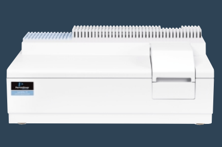
UV-VIS SPECTROPHOTOMETER WITH DRS (Under Installation)
Make: PerkinElmer, Model: Lambda 365+
UV-VIS SPECTROPHOTOMETER WITH DRS diffuse reflectance spectroscopy and can be applied to measure the absorption of the sample surface and interior by the reflection of light. It can also be used to analyze the structure and composition of the opaque material i.e. physico-chemical study of surfaces.. This equipment has wide application in various fields such as such as color measurements of textiles, pharmaceuticals, building materials, paper and pulp materials etc., to adsorption studies and other basic investigations in physical, inorganic and organic chemistry.
INSTRUMENT IN PIPELINES
- Stereozoom microscope
- Petrological microscope
- Cryomicrotome
- Dual block PCR
- Real Time Quantitative PCR
- Lyophiliser
- Protein Purification system
- Orbital Shaker cum Incubator
- FT-IR with ATR
- Plant Growth chamber
- Solar Simulator
- Differential Global Positioning system
In addition, the following facilities are developed in the CIF for the Centre of Excellence in Integrated Omics and Computational Biology with financial assistance from WB-OHEPEE
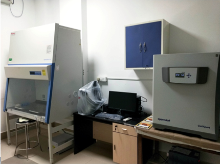
CELL CULTURE FACILITY
The facility provides necessary infrastructure for culturing and maintaining cell lines (mammalian) for experimental purpose. The facility is equipped with Biosafety cabinet (Make: Thermo Fisher Scientific), Inverted Microscope (Make: Leica Microsystems), CO2 Incubator (Make: Eppendorf) and all the accessory small instruments.
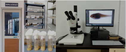
DROSOPHILA CULTURE FACILITY
The facility is a collection of commonly used, genetically defined Drosophila melanogaster, an insect which is extensively used as a model organism to address a wide range of biological questions and their validations. The facility is equipped with a Stereozoom microscope (Make: Leica Microsystems) and all necessary accessories for the maintenance of the cultures.
CLONING FACILITY
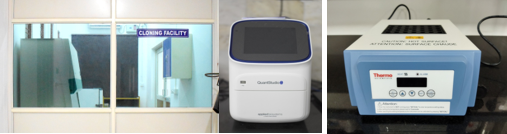
The molecular cloning facility is developed to analyse qPCR of DNA and RNA samples, plasmid clones, and molecular biology related experiments. The facility is equipped with Laminar hoods (Make: Mikrofilt), Real Time PCR (Make: Applied Biosystems), Gradient PCR (Make: Bio-Rad), Dry bath (Make: Thermo Fisher Scientific), Gel electrophoresis system (Make: Bio-Rad), and accessory small instruments.
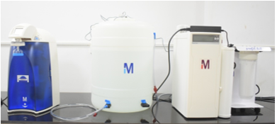
WATER PURIFICATION SYSTEM (MAKE: MERCK MILIPORE)
The facility provides ultrapure water for experimental purpose by removing ionic and organic contaminants, particulates, bacteria, pyrogens, nucleases, proteases, VOCs, endocrine disruptors and organics for LC and others.
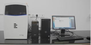
GEL DOCUMENTATION SYSTEM (MAKE: VILBER)
The equipment is used to record and analyze the results of gel electrophoresis and membrane blotting experiments. It is highly essential for visualizing stained or labeled nucleic acids and proteins in agarose, acrylamide or cellulose.
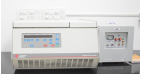
REFRIGERATED HIGH-SPEED CENTRIFUGE (MAKE:BECKMAN COULTER)
The equipment has wide range of volumetric capacity and is used in cell culture and bacteria collection, concentration, ad lysate collection and purification.
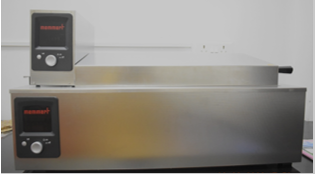
Shaking Water Bath (Make: Memmert)
The equipment has precise temperature control and a smooth reciprocal horizontal shaking motion. It is ideal for thawing, heating, mixing and shaking samples. It is mostly used for molecular and biochemical experiments.
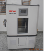
ORBITAL SHAKING INCUBATOR (MAKE: REMI)
The facility is designed for precise temperature control with simultaneous shaking option, mostly used in microbiology, fermentation, tissue culture and other biotechnology experiments.